

A baby’s heart starts forming very early on, and its development matters a lot for how the rest of the pregnancy goes. In some cases, doctors will ask for a fetal echocardiography, which is basically a specialised ultrasound that takes a much closer look at the baby’s heart before delivery. The idea is to catch heart abnormalities early, so if something needs attention, there’s time to plan for it.
What Is Fetal Echocardiography?
A fetal echocardiography, or a fetal echo scan, is a detailed ultrasound scan focused entirely on the baby’s heart, how it’s structured and how it’s working.
A normal pregnancy ultrasound checks the whole baby. This one is different. It zooms in specifically on the heart chambers, valves, major blood vessels, blood flow, and heartbeat. Most of the time it’s done in the second trimester, somewhere between 18 and 24 weeks, though that window can shift a bit depending on why the test was ordered in the first place.
Why Is Fetal Echocardiography Done?
There are a handful of reasons a doctor might recommend this test. Usually it comes down to a higher-than-average risk of a congenital heart defect. That can include things like:
- A family history of congenital heart disease
- Something that looked off on an earlier routine ultrasound
- Certain health conditions or infections in the mother
- Genetic or chromosomal conditions found during pregnancy
- Other abnormalities spotted elsewhere in the baby’s body
- Medications or exposures during pregnancy that could raise the risk
- A previous pregnancy where the baby had a heart defect
Just because a fetal echo scan is recommended doesn’t mean anything is actually wrong. A lot of the time, the test just confirms the heart looks the way it should.
How Is the Procedure Performed?
It’s a straightforward ultrasound, nothing surgical, no needles involved.
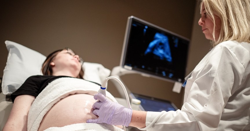
The mother lies down on the exam table, and gel gets applied to the belly. Then a transducer is slowly moved over the abdomen, and the sound waves it picks up produce images of the baby’s heart on screen. From there, the specialist looks closely at the structure, movement, and flow of blood through it.
It doesn’t hurt. Depending on how the baby is positioned and how much needs to be checked, it can take anywhere from half an hour to an hour.
Benefits of Fetal Echocardiography
The biggest upside is simply getting a clearer, earlier picture of the baby’s heart. That can mean:
- Certain congenital heart defects get caught before birth
- Doctors get a better sense of how the heart is actually functioning
- It becomes clearer whether more monitoring is needed
- Parents and doctors have time to prepare if specialised care is going to be needed after delivery
- Delivery and newborn care can be planned around more serious heart conditions ahead of time
Are There Any Risks?
This test relies on ultrasound, not radiation or X-rays, so when it’s carried out by a trained professional and for a genuine medical reason, it’s considered safe.
Still, no ultrasound catches everything. Some heart defects are too small to see before birth. Others don’t really show up until later in the pregnancy. And the accuracy of the scan can depend on things like the baby’s position, how far along the pregnancy is, and other technical factors on the day.
Final Thoughts
Fetal echocardiography gives a much closer look at a baby’s heart than a standard scan can. If a doctor recommends it, it’s worth going ahead with, since it gives real insight into how the heart is developing and helps everyone involved prepare for whatever care might be needed, before or after the baby arrives.
If you’re looking for a place to get this done, Apollo Cradle & Children’s Hospital, a leading fetal medicine centre in Indiranagar, Bangalore, is a good option to consider. It gives you access to specialised care and detailed fetal heart assessment under the guidance of experienced medical professionals.
 Cares My Health Blog